Inspiration #1
Training in Otology has included training on cadaveric temporal bones for over 100 years. Otologists recognized that the anatomy they work with in the ear is full of essential structures that are closely juxtaposed next to one another. As such it is easy to injur those structures making ex-vivo training that much more important. We agree that having a detailed knowledge of anatomy makes operating on critical structures safer, faster and more effective.
Inspiration #2
In 2001-2002, Dr. Steven Zeitels, my laryngology fellowship director at the time, was in the process of introducing new surgical techniques for voice restoration, especially for unilateral vocal fold paralysis (1, 2 – see references below). These techniques introduced concepts that were difficult to fully grasp without understanding the laryngeal anatomy and physically manipulating the component structures freely. Obviously, there are limitations to learning this in the living and a need for an ex-vivo opportunity was born.
First Steps
With the assistance of Dr. James Kobler, PhD at the Massachusetts Eye and Ear Infirmary, we designed and built a physical station (made out of plywood, steel and brass) designed to allow the use of cadaveric larynges for simulated endoscopic and open laryngeal surgery (3). This station allowed me and subsequently many others to practice laryngeal procedures and even to conceive of new ones (4). Central benefits of practicing on cadaveric larynges are that the anatomy is either identical in the case of human larynges or reasonably close in the case of canine larynges or even porcine larynges. Also the tissues subtypes such as cartilage are identical and help the learner to learn anatomical relationships and tissue tolerances in a zero-risk environment. The station is also such that it can be used for solo work or with one or more people simultaneously. When a microscope is used for endoscopic work the microscope usually has a “teaching head” so that another person can watch in real time; modern microscopes almost all have video output so that the surgeon’s view is displayed on a video monitor for group learning.
 Codifying the Learning

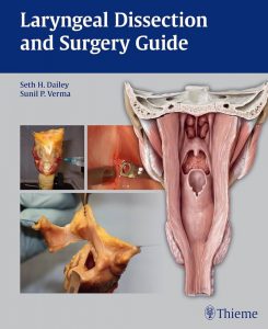
With the encouragement of our first laryngology fellow at the University of Wisconsin-Madison, Dr. Sunil P. Verma, a laryngeal surgery guide was pursued. He and I asked expert authors to contribute chapters on 33 very specific procedures. Each chapter was illustrated with the laryngeal dissection station and cadaveric larynges to illuminate ideas. Each chapter has had key clinical points, pearls and pitfalls and appropriate references. Dr. Verma and I and our co-authors finished the book and it was published by Thieme in 2011. It was modified and translated into Mandarin in 2013.
Refinements
Dr. Verma introduced a variant of the original station to create an alternative low-lost physical platform for laryngeal surgical learning. He and his team called this VOCALSS: Versatile optimally constructed aid for laryngeal surgery simulation (5).
Regional Contributions and the Laryngeal Dissection Course
The above-mentioned book grew out of efforts to educate our UW Otolaryngology-Head and Neck Surgery residents on laryngeal anatomy and surgery. Initial efforts were in person and hands-on and took place in the temporal bone lab. We performed numerous standard procedures such as vocal fold injection, epithelial vocal fold resection, thyroplasty and variants of laryngoplasty. Teaching aids were chalkboards and stapled together surgical guides!
Eventually the stapled surgical guides became the above referenced book which we now use as our main teaching guide. Recognizing the value of community and diversity of perspectives, we then began to invite regional laryngology faculty and their associated Otolaryngy-Head and Neck surgery residents to participate in the course. These have included faculty and residents from Loyola, Rush, University of Chicago, Northwestern, Medical College of Wisconsin, University of Iowa, Mayo-Rochester, Cleveland Clinic, Case Western and others. We deeply appreciate the generosity and selflessness of all of the faculty who hav contributed over the years. We would like to specially recognize Dr. Joel Blumin of the Medical College of Wisconin who has donated his time and teaching to ALL 16 Laryngeal Dissection Courses that we have held – thank you, Joel, all.
We have had the good fortune of having additional faculty from the US join us as well. The course was a CME approved course briefly. Then the worldwide COVID-19 pandemic happened and the course had to change. We reverted to a UW residents hands on course initially. As we re-emerged from the pandemic, the notion of introducing in-person Master Classes came to be; a surgical expert would show small groups how to perform an operation using the cadaveric platform for demonstration. This offering was instantly recognized as being a high-yield learning opportunity. So, as all things involving COVID, it became a virtual offering.
For the last few years we have offered a half day session of 6 Master Classes in the morning and a hands-on session in the afternoon for local and regional residents and faculty in our beautiful Simulation Center. For the Master Classes we try to have a balanced program of operations/procedures that pertain to voice, swallowing and airway with a principal focus on the adult world but also some pediatric laryngology options. Each Master Class is a pre-recorded video with over narration by the presenter. In their video they demonstrate an operation/procedure with particular emphasis on the key pearls and pitfalls. After the video the presenter is available for a LIVE Question and Answer session.
We have had up to 400 participants from over 15 countries and 4 continents sign up for participate in this course and we are still growing. Forward!
References
- Adduction arytenopexy: a new procedure for paralytic dysphonia with implications for implant medialization.
Zeitels SM, Hochman I, Hillman RE.Ann Otol Rhinol Laryngol Suppl. 1998 Sep;173:2-24
PMID: 9750545 - Cricothyroid subluxation: a new innovation for enhancing the voice with laryngoplastic phonosurgery.
Zeitels SM, Hillman RE, Desloge RB, Bunting GA.Ann Otol Rhinol Laryngol. 1999 Dec;108(12):1126-31. doi: 10.1177/000348949910801206.
PMID: 10605916 - A laryngeal dissection station: educational paradigms in phonosurgery.
Dailey SH, Kobler JB, Zeitels SM.Laryngoscope. 2004 May;114(5):878-82. doi: 10.1097/00005537-200405000-00017.
PMID: 15126748 - Local vascularized flaps for augmentation of Reinke’s space.
Dailey SH, Gunderson M, Chan R, Torrealba J, Kimura M, Welham NV.Laryngoscope. 2011 Feb;121 Suppl 3(Suppl 3):S37-60. doi: 10.1002/lary.21186.
PMID: 21271606 - VOCALSS: Versatile optimally constructed aid for laryngeal surgery simulation.
Foulad A, Bui P, Dailey SH, Verma SP.Laryngoscope. 2015 May;125(5):1169-71. doi: 10.1002/lary.25091. Epub 2014 Dec 24.
PMID: 25545359